What is mass cytometry?
Mass cytometry is a laboratory method that allows researchers to analyze individual cells and their biological characteristics. Using antibodies tagged with rare earth metals, it can measure a wide range of features on and inside cells, such as whether a cell is activated, where it may migrate in the body, or which signaling molecules it produces. In practice, around 60 different parameters can currently be measured per cell.
What is it used for?
This information helps researchers better understand how cells function in health and disease, and where these processes go wrong in blood or tissue. It can also be used to identify biomarkers that may predict whether a patient will respond to a particular treatment. Because mass cytometry can analyze large numbers of cells at once, even rare cell populations can be captured effectively.
How does mass cytometry work?
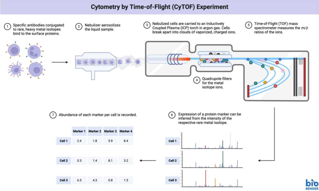
Mass cytometry combines principles of flow cytometry with inductively coupled plasma mass spectrometry (ICP-MS), a technology originally developed for trace element analysis. First, cells are isolated from blood or tissue and stained with antibodies that bind specifically to structures inside or on the cell surface. These structures may include receptors, enzymes, or signaling molecules that are relevant to a biological process or disease.
The antibodies are labeled with rare earth metals, which can be detected by the mass cytometer. Compared with the fluorescent dyes used in conventional flow cytometry, this approach makes it possible to measure many more markers at the same time, generating detailed information even from very small sample volumes.
The labeled cells are then introduced into the mass cytometer. There, they are broken down into individual atoms by an argon plasma heated to approximately 5,000 °C. Unwanted elements such as oxygen, hydrogen, and nitrogen are removed, leaving the rare earth metals for detection. Each cell’s atomic signal is then analyzed in a time-of-flight mass spectrometer according to mass and flight time. This creates a profile for every single cell, showing which markers were detected and, therefore, what characteristics the cell has.
A newer development is Imaging Mass Cytometry (IMC), an extension of conventional mass cytometry. In IMC, a laser ablates the tissue sample spot by spot, and the released material is analyzed by mass spectrometry. This preserves the spatial position of each cell within the tissue while measuring its biological properties. Researchers can therefore analyze spatial information such as cell arrangement in tissue, neighboring cells, and cell–cell interactions, combining this with the high-parameter capabilities of mass cytometry.